Monday, December 26, 2011

Journal paper: Choroidal optical coherence angiography with variable velocity range

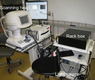
Non-invasive and three-dimensional angiography of choroid with several blood-velocity range is available now. Our colleague Franck Jaillon recently published the details of this methodology in Optics Express. In this paper he demonstrated non-invasive visualization of fine vasculature of the choroid of normal eyes. This technology is based on Doppler optical coherence tomography, and hence it is totally noninvasive and three-dimensional. The Doppler detection is performed with our original dual beam scan Doppler scheme, which provides extremely high Doppler sensitivity. In addition, this system uses a 1-um probe beam, it provides deep penetration into the choroid.

   The details are described in the following paper.
>> Full length article on Journal web-site (open access)
Citation: F. Jaillon, S. Makita, Y. Yasuno, "Variable velocity range imaging of the choroid with dual-beam optical coherence angiography," Opt. Express 20, 385-396 (2012).

Tuesday, July 26, 2011

Journal Article: Office-Based Polarization Sensitive OCT for Anterior Eye

Our colleague Yiheng Lim recently reported a clinical prototype of polarization sensitive optical coherence tomography (PS-OCT) specially designed for the investigation of anterior eye segments. This system based on our developed high-speed Jones matrix OCT methodology, and integrated with a scanning head specially designed for anterior eye imaging. This clinical prototype provides semi-automatic alignment function and possesses a compact, office-compatible size. Several clinical cases were investigated to prove the clinical utility.

The details are reported in Biomedical Optics Express.

>> Full length article on journal web-site (open access)

Citation: Y.H. Lim, M. Yamanari, S. Fukuda, Y. Kaji, T. Kiuchi, M. Miura, T. Oshika, and Y. Yasuno "Birefringence measurement of cornea and anterior segment by office-based polarization-sensitive optical coherence tomography," Biomed. Opt. Express 2, 2392-2402 (2011).

Sunday, April 10, 2011

Journal Article: High-Sensitive Doppler Imaging of Choroid by Dual-Beam Doppler OCT

Our colleague Franck Jaillon recently reported his new Doppler optical coherence tomography (OCT) system which enables high-penetration and dual-velocity imaging of choroidal vasculature. This methodology employed   newly developed dual-beam OCT scanning head to provide high-sensitive Doppler imaging. Furthermore, this scanning head can provide multiple velocity range for Doppler detection. Since this system is using 1-um light as a probe, the details of in vivo choroidal vasculature was successfully visualized.

The details are published in Biomedical Optics Express.

>> Full length article on journal web-site (open access)

Citation: F. Jaillon, S. Makita, E.-J. Min, B.H. Lee, Y. Yasuno, "Enhanced imaging of choroidal vasculature by high-penetration and dual-velocity optical coherence tomography," Biomed. Opt. Express 2, 1147-1158 (2011).